The Etching That Revealed a Time Capsule
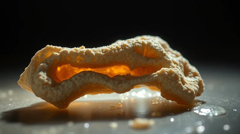
The fossil that produced this result would never have been chosen for a museum wall. It is a nondescript fragment of hind-limb bone from a theropod dinosaur called Aratasaurus museunacionali, recovered in northeastern Brazil in 2008. Kliti Grice, a professor at Curtin University in Perth, Australia, describes the material plainly: “The fossils we worked on are not display specimens by any means, so they don’t look very interesting. If you look at them, you think there’s just a bunch of bones and there’s probably not much in there.” [2] That judgment — bones, nothing more — is exactly what the new work overturns.
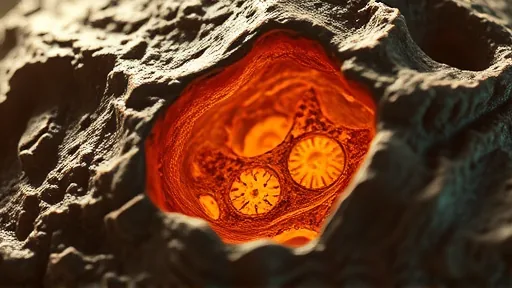
The method behind the reversal is almost aggressively simple. Grice’s team etched away part of the fossil with hydrochloric acid, dissolving the surrounding rock to expose what lay inside. What she saw sent her running: “I was jumping up and down. I ran up to my head of school’s office and was so excited that he gave me a whisky to calm me down. No one has ever seen dinosaur red blood cells in high resolution before.” [2] The cells were not scarce. In a single slide just a fifth of a millimetre wide, the team counted 20 blood cells. Every one of them sat inside vascular canals — the channels that once carried blood vessels through the bone — which is precisely where red blood cells belong and precisely where a contaminant would be least likely to land. The location is itself part of the evidence.
What separates this find from earlier claims is chemistry performed in place. Using high-performance liquid chromatography–mass spectrometry, the team identified parent and diagenetically altered heme B as the principal tetrapyrroles in the bone extract. [1] Heme B is the oxygen-binding component of haemoglobin, the molecule that makes blood red and carries oxygen through a living body. The surrounding sediment told the opposite story: it was dominated by chlorophyll and bacteriochlorophyll porphyrin signatures, the chemistry of algae, not of blood. Two different chemical worlds, one inside the bone and one outside it.
The team then confirmed the link between chemistry and structure using spatially resolved analyses. Raman spectroscopy and transmission electron microscopy showed that the porphyrin signals of heme and the key elements of heme — iron, nitrogen and carbon — were localised to the ellipsoid microstructures themselves. [1] Transmission electron microscopy also detected hausmannite, a manganese oxide with the formula Mn3O4, a mineral the team describes as a natural stabiliser that helped preserve the cellular structures and biomolecular components across geological time.
Old Drawers, New Questions
The scale of this result is best measured against what came before. Red blood cells had already been reported in dinosaur remains: bone fragments collected in Canada in 2015, dated to 75 million years ago, and the femur of a Tyrannosaurus rex that died 68 million years ago, found in Montana in 2005. Each of those finds pushed the boundary back by tens of millions of years. The Aratasaurus material is dated 40 million years earlier than the Canadian find and, more importantly, changes what counts as proof. Previous studies reported possible red blood cell structures. This study is the first to chemically demonstrate haemoglobin-derived molecules inside the cells themselves.
The individual studied was not fully grown. Its estimated body length was just over 3 metres and its weight about 34 kilograms — a juvenile theropod, the group of two-legged, mostly meat-eating dinosaurs that included Tyrannosaurus rex and Velociraptor. When the researchers compared the fossil cells with those of the dinosaur’s closest living relatives, birds and crocodiles, the Aratasaurus cells were significantly larger. That gap has two possible readings, and the team offers both without choosing: the cells may have swollen during fossilisation, or the species may have grown far larger than this particular specimen did.
Grice draws a practical consequence that reaches into every museum storeroom on earth. “There’s drawers and drawers of fossil material, which don’t look very nice,” she says. “This kind of work opens up a whole new opportunity to unlock those collections and bring modern technology and molecular techniques to these specimens.” The logic is uncomfortable for museum collections: the specimens judged least photogenic may be the ones holding the most information, and nobody has yet looked at them with these tools.
The team believes the exceptional preservation resulted from the dinosaur being buried rapidly in a low-oxygen, microbially rich environment shortly after death. Grice frames the implication directly: “These findings challenge longstanding assumptions that certain biomolecules such as haemoglobin cannot survive deep time.” [2] Martin Van Kranendonk, head of Curtin’s School of Earth and Planetary Sciences, extends the point beyond palaeontology, noting that the research redefines how molecular structures can be preserved over long time frames or in hostile conditions, with possible implications for astrobiology and the search for ancient life in the cosmos.
The Shape That Does Not Quite Fit
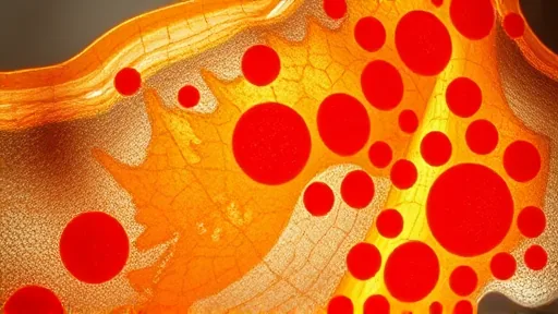
Not everyone accepts the conclusion. Some experts argue that the shape of the structures does not match reptilian red blood cells — a morphological objection that chemistry alone cannot settle. The team’s answer is that it did not rely on chemistry alone. As Grice puts it: “We combined microscopy, mineralogical analyses and molecular geochemistry. Critically, we investigated the chemistry in situ, so we could relate the chemical signals to the microscopic structures in their physical context within the fossil. That is much stronger than simply extracting molecules from a piece of fossil and assuming where they originated.” [1] That defence sharpens rather than resolves the dispute. The in-situ approach ties molecules to structures, but it cannot tell a critic whether the structures are red blood cells that swelled, cells of a different type entirely, or mineral casts that mimic a cell’s outline. The size discrepancy against birds and crocodiles sits at the centre of this: it is either evidence of fossilisation damage or evidence that the identification is wrong, and the same observation supports both readings. A 115-million-year-old argument now rests on the outline of ellipsoidal microstructures, each smaller than a fifth of a millimetre, and the question of what shape a dinosaur’s blood cell was supposed to have.