One Flash, Twenty Meters per Second
The parameter that governs this entire image is time, measured in fractions of a second. An inkjet nozzle fires a droplet that crosses the gap to the page at roughly 20 meters per second — fast enough that even the best slow-motion camera cannot resolve the moment. Everything else in the setup is ordinary: a printer, a sheet of paper, a microscope. Only the exposure is extraordinary, and it is the single variable that decides whether the picture exists at all.
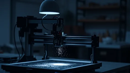
Ole Bielfeldt, a design student in Cologne, Germany, solved the problem by inverting the machine’s logic. Instead of holding the paper still and moving the print head, he built a rig that moves the paper while the ink nozzles stay fixed and in focus. The contraption is only a bit larger than his head — a small workshop object that produces a picture of something most people never consider.
Then came the darkness. Bielfeldt worked with the lights off and lit the scene with one brief flash, a duration short enough to stop the droplet mid-flight. He instructed the printer to spell the word “macro,” a nod to his social media handle, macrofying, and repeated the run until the timing landed. The result took first place in the 2026 Nikon Small World photomicrography competition. [1] The path to that rig began as a quarantine hobby. Around 2021, an old school microscope from his attic gave Bielfeldt a way to explore while stuck at home during the pandemic. Before the project, he says, a printer was “like magic.” Photographing its interior turned the magic into mechanism. “It’s so, so advanced technically, but at the same time it’s so, so common,” he says.
A Parallel Line of Inquiry in Helsinki
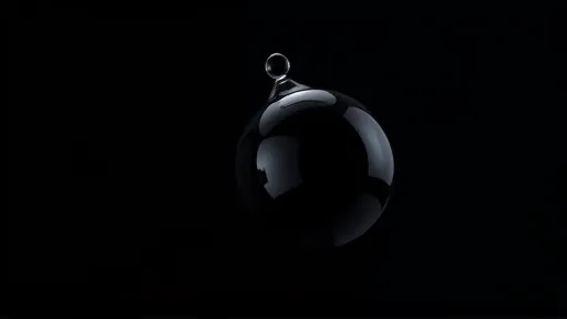
A second group works the same problem from the opposite direction: not freezing motion in air, but mapping structure inside tissue. Cell biologists Sinem Karaman and Satu Paavonsalo at the University of Helsinki visualize vascular networks — the branching systems of blood vessels — to study how they function within tissues.
Their subject is a mouse eye, captured as a three-dimensional cross section rather than the flattened retinas her lab typically uses. The image is crisscrossed with fractal branches of blood vessels and tethered by the optic nerve. Getting there required meticulous mounting across several attempts before Paavonsalo was satisfied with the arrangement. Even then, certainty was out of reach. “I had to hope that the sample was correctly positioned and that the staining was detected throughout the sample,” she says. Her excitement on seeing the finished image was, by her account, all the more pronounced for it. The photograph placed 19th in the competition. [1] The arrangement itself carries a research question. Paavonsalo says the pattern of vessels could point to mechanisms underlying how these systems stop working in obesity and diabetes.
Other Winners in the Same Contest
The competition recognized 86 images in total, announced October 21, and several of the remaining honorees show the same discipline of preparation applied to living subjects. [1] John-Oliver Dum, whose fascination with spiders began as a childhood fear after a painful bite, turned to studying the movements of a curly hair tarantula, Tliltocatl albopilosus. His photograph of a pollen-dusted jumping spider took second place. The logistics stretch across seasons: his wife grows plants selected for their pollen, which he uses in his images. “The biggest challenge was finding a spider like this on the right plants, with the right pollen attached to it, at exactly the right time of year and at exactly the right moment,” he says. “You need to understand not only the spider’s behavior, but also the surrounding insect and plant life and their seasonal rhythms.” His hope is that the picture makes viewers look more closely: “We first have to see the beauty of this largely unknown world before we can truly understand what we may be destroying.”
At the University of Montpellier in France, evolutionary developmental biologist Gonzalo Quiroga-Artigas captured a tardigrade, Hypsibius exemplaris, emerging from its shed cuticle with an egg left behind — an animal only about two-and-a-half times as wide as a strand of hair, and one able to survive space by entering suspended animation. The timing is the difficulty. “Egg laying is closely synchronized with molting,” he says, and the moment of shedding passes quickly enough to be easy to miss. A tardigrade can lay up to about a dozen eggs at once. He chose this specimen, which placed 12th, because “the egg looks almost like a precious gem being left behind by its mother.” In Brooklyn, Eric Epstein found a slime mold, Hemitrichia sp., sprouting from a decaying log in Prospect Park. He cut pieces of waterlogged wood free without disturbing his subjects — “in this liquidy state, any touch would demolish” the fruiting bodies — and stacked 240 images to bring the gelatinous structures into sharp detail. The photograph placed sixth. Slime molds have already earned a place in research: one species closely mirrors the cosmic web of gas and dark matter connecting galaxies, helping scientists map the cosmos more accurately, and another has fascinated researchers for packing a store of bacteria to feed the next generation when it is near death.
At the University of California, Berkeley, undergraduate student Faith Fernandes and structural cell biologist James Glover produced an image of human induced pluripotent stem cells that had developed into neurons — cells reprogrammed to a stage before they were locked into a specific fate. Clusters of nuclei look like flashpoints in an intricate web; the neurotubules that give the cell structure and transport molecules within it appear blue, and orange marks where the DNA sits. Fernandes made the image her computer wallpaper as soon as it was taken. She says she hopes studying these cells will illuminate ways to address neurodegeneration in diseases such as Parkinson’s disease. Researchers have already used such cells to regrow eye tissue, to address premature aging in mouse studies, and to grow organs. The shot earned an Image of Distinction title.