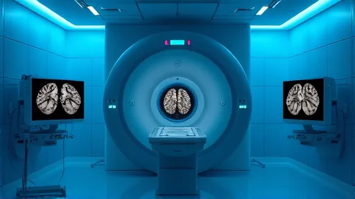
A radiologist stares at a stack of two-dimensional MRI slices, each one meant to be a thin horizontal cut through a fetal brain. If the fetus held still, those slices would line up neatly and reconstruct into a clean three-dimensional volume. But fetuses move. Each slice may come from a slightly different angle, and the resulting gaps can hide the very regions a physician needs to see. The judgment call — is this a normal brain or an abnormality — depends on an image that may simply be incomplete.
The problem with a moving target
Fetal MRI begins with a routine prenatal ultrasound. When a sonographer flags a possible brain abnormality, the patient is referred for magnetic resonance imaging, which offers finer detail than ultrasound can. The difficulty is that the scan takes time, and the subject does not cooperate. Ramya Muthukrishnan, a PhD student in the lab of Polina Golland at MIT, described the core issue in plain terms: “One of the problems with having the baby move is that the slices are all over the place.” [1] A series of 2D images taken during the scan are supposed to stack into one high-resolution 3D picture of the fetal head. When the head shifts, each slice can be captured from a different orientation, producing coverage gaps and unusual slice angles. The physician reviewing the result may miss a pathology or misread one.
The stakes are not abstract. A pregnant mother can spend a long time in the scanner and leave without a usable image, uncomfortable and unresolved. The radiologist, meanwhile, has no complete volume to interpret. Watching that frustration is what set her research agenda: the gap between what the technology could capture and what the patient and physician actually received.
What the model does differently
Researchers at MIT, Boston Children’s Hospital, and Inria built and trained a deep learning model called E(3)-Pose to address this. [1] The model helps image the fetal head while it is in motion, tracking orientation well enough to assemble a high-resolution volume a radiologist can examine. A paper on the work was presented at the 2026 European Conference on Computer Vision.
The design choice that distinguishes E(3)-Pose is architectural rather than purely empirical. Instead of training a network to infer physics from data alone, the researchers embedded physical principles directly into the model. Left-right symmetry of the fetal head is built into the network’s structure, which makes it easier for the model to identify the brain’s orientation correctly. This is the difference between teaching a system to recognize a pattern and giving it a prior that constrains what it can conclude. The symmetry assumption is not learned; it is assumed, and the learning happens within that constraint.
The gain here is concrete and narrow: better tracking of a moving object so that scattered 2D slices can be assembled into a usable 3D image. It does not diagnose anything. It does not replace the radiologist. It improves the input that human judgment depends on, which is precisely where the current process fails.
From the scanner to the clinic
Translating a model from a GPU to a working MRI scanner turned out to be its own ordeal. When Muthukrishnan began the project at the start of her PhD, she had no background in physics, let alone MRI physics. Most of her experience was in computer science. The gap between a promising architecture and a functioning clinical tool is measured in troubleshooting sessions, not benchmark scores.
That gap was bridged partly through collaboration. The work was guided by senior authors Elfar Adalsteinsson, MIT professor of electrical engineering and computer science; Ellen Grant, the Käthe Beutler Professor of Pediatrics and professor of radiology at Harvard Medical School; and Golland. [1] Both point at the same thing: the model’s value is measured in what it hands to the clinician, not in what it claims on its own.
Where the gain stops
The earlier a brain abnormality is detected, the easier it is to intervene, which makes the second trimester the more consequential target and the harder technical problem. The current version does not reach it. tonomous driving, where limited sensors must capture noisy 3D data to track objects in three dimensions. Those are plausible extensions, not demonstrated results. The honest summary of E(3)-Pose in 2026 is a model that improves motion tracking for a specific and difficult imaging problem, built on a physics-informed architecture, validated in a clinical research setting, and still limited to the fetal population that is easiest to image and latest to benefit from early detection. The promise of earlier intervention remains on the other side of the second-trimester gap.