AI Bubble Detection Lacks Precision in Cardiac Surgery
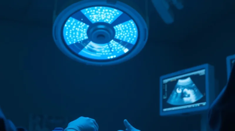
The operating room has gone quiet in a way that has nothing to do with sound. Monitors hum, ventilators cycle, and the surgical team moves with the choreography of people who have done this a thousand times. But the quietest presence in the room is the algorithm — the one that watches the ultrasound feed, the one that promises to see what human eyes cannot. In cardiac surgery, where a stream of microscopic air bubbles can slip into the bloodstream and travel toward the brain, that promise carries unusual weight. A gaseous microembolus is not a dramatic event. It does not announce itself. It simply arrives, and the damage it does accumulates silently across a lifetime of small insults.
The Problem with Seeing Everything
For decades, surgeons have relied on transesophageal echocardiography to monitor these bubbles in real time. The technique is elegant in principle: a probe placed in the esophagus provides a window into the beating heart, and the clinician watches for the bright specks that indicate circulating gas. The reality is messier. The heart moves, the probe angle shifts, and the background is crowded with structures that look almost exactly like the thing you are trying to find. A skilled echocardiographer develops an eye for it, but that eye is a scarce resource, and it tires. The problem was never that the data was missing. The problem was that the interpretation asked too much of the people doing it.
This is where the machine enters, carrying the familiar promise of relief. The research team behind this study proposed a 2.5D U-Net architecture — a deep learning model designed to detect these bubbles in the space-time volume of ultrasound data, rather than in individual frames. The idea has an intuitive appeal that is hard to resist. If a human expert can learn to spot the pattern of a moving bubble across consecutive frames, why should a machine not learn it faster, and do it without blinking? The model was trained on eight recordings from eight different patients, each undergoing cardiac surgery, and the results looked strong on paper. Precision of 92.55 percent, recall of 80.54 percent, inference time of 0.12 seconds per batch. Real-time performance, the authors concluded, is technically feasible.
What the Numbers Don’t Say
But here is where the careful reader must slow down, because the paper contains a confession buried in its own metrics. The researchers evaluated their model under two different scoring systems. The first was generous: a “radius-tolerant” grace zone that counted a detection as correct if it landed within three pixels of the true bubble location. Under this system, the model looked excellent. The second evaluation was strict: pixel-by-pixel segmentation, where every single pixel had to match perfectly. Under this system, the model’s performance collapsed. The intersection over union — a standard measure of how precisely the model identifies the target region — fell from 73.95 percent to 41.74 percent. The Dice coefficient fell from 84.13 percent to 57.98 percent.
The gap between these numbers is not a technical footnote. It is the whole story. The model is very good at pointing in the general direction of a bubble. It is considerably less good at telling you exactly where the bubble is. For a screening tool, that might be acceptable. For a clinical decision support system in the middle of a cardiac procedure, the difference between “somewhere around here” and “precisely here” is the difference between reassurance and truth. The grace zone did not simply make the model look better. It changed the nature of what the model was claiming to do.
The Comfort of Approximate Truth
There is a pattern here that extends far beyond cardiac surgery. We have become accustomed to AI systems that perform brilliantly under evaluation conditions and then reveal their limitations when the conditions change. The mechanism is not deception in the malicious sense — the algorithm is not lying to anyone. The deception is structural, embedded in the way we choose to measure performance. When we design the evaluation to accommodate the model’s weaknesses, we are not testing the model anymore. We are testing our own tolerance for imprecision.
The researchers did include one additional check that deserves attention. They ran the model on an external dataset of TEE recordings that contained no bubbles at all, to see whether it would produce false alarms. The model produced mostly empty masks, indicating a low rate of spurious detections. This is genuinely reassuring, and it is the kind of test that should be standard practice everywhere. But it also highlights how unusual this level of rigor still is. Many AI papers in medicine report only the optimistic numbers, the ones that come from the comfortable evaluation conditions, and never ask what happens when the model encounters a world it has not been prepared for.
The Weight of the Empty Mask
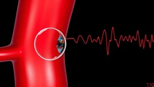
Think about what an empty mask means in this context. The model looked at a heart that was clean, and it said so. That is the easy part. The hard part is the near-miss, the bubble that was there but too small, too fast, too obscured by the surrounding tissue. The strict metrics suggest that the model misses a meaningful fraction of these. In a clinical setting, a missed bubble is not a statistic. It is a potential stroke, a cognitive decline that shows up years later, a family member who is not quite the same after surgery. The model does not feel the weight of that. The surgeon does.
The study’s authors are careful in their language. They call it a feasibility study, and they are right to do so. Eight patients is a pilot dataset, not a proof. The leave-one-patient-out cross-validation is a reasonable approach for such a small sample, but it cannot capture the full variability of human anatomy, the range of ultrasound machines, the differences in clinical conditions, or the many other factors that could affect the model’s performance in a real-world setting. The model works on the data it was given. Whether it works on the next patient, in the next operating room, with the next ultrasound technician’s particular way of holding the probe — that remains an open question.
The Architecture of Trust
There is a deeper issue lurking beneath the technical details, and it has to do with how we build trust in systems we do not fully understand. The 2.5D U-Net is not a simple algorithm. It is a deep neural network with millions of parameters, trained through a process that even its creators cannot fully trace. When it makes a mistake, it does not make the kind of mistake a human makes. A human echocardiographer who misses a bubble can often explain why — the angle was bad, the image was noisy, the patient moved. The model cannot explain anything. It simply produces an output, and the output is either accepted or not.
This is the moment where the concept of AI deception becomes genuinely important. Not deception as intentional falsehood, but deception as a gap between appearance and reality. The model appears to be detecting bubbles. It produces masks that look like the masks a human expert would produce. But the process by which it arrives at those masks is fundamentally different, and the difference matters when things go wrong. A human can be questioned. A model can only be re-run.
The Quiet Shift in Responsibility
Consider what happens to the surgeon’s attention when such a system is deployed. The algorithm is fast, it is always on, and it produces a constant stream of visual output. The temptation is to let it become the primary observer, with the human reduced to a supervisory role. This is not a hypothetical concern. It is the trajectory that every AI-assisted medical technology has followed since the first computer-aided detection systems were introduced in radiology decades ago. The human does not stop watching, but the watching changes. It becomes a monitoring of the monitor, a second-order attention that is not the same as the first-order attention required to actually see the bubble.
The study’s inference time of 0.12 seconds per batch is impressive, and it is the kind of number that gets quoted in press releases. But real-time performance is not the same as real-time understanding. The model can process the data fast enough to keep up with the ultrasound feed. That does not mean it understands what it is seeing in the way a clinician understands it. The distinction sounds philosophical, but it has practical consequences. When the model is wrong, the error is not just a wrong answer. It is a wrong answer delivered with the same confidence as a right one, and that confidence is contagious.
The Limits of the Grace Zone
Let us return to that three-pixel grace zone for a moment, because it deserves closer scrutiny. In an image of 600 by 800 pixels, three pixels is a tiny distance. But the bubbles the model is trying to detect are also tiny, often just a few pixels across. A three-pixel tolerance is not a minor adjustment. It is a fundamental redefinition of what counts as success. Under the strict metric, the model’s precision and recall were dramatically lower, and while the authors do not report those numbers in the abstract, the IoU and Dice coefficients tell the story clearly. The model is not precisely locating the bubbles. It is locating the general area of the bubbles, and the grace zone was designed to make that look like precision.
This is not an accusation of bad faith. The researchers are transparent about their methodology, and the inclusion of strict metrics is a sign of intellectual honesty. But the choice of the grace zone as the primary evaluation criterion reveals a preference for optimism over accuracy, and that preference is widespread in the field. We want AI to work, so we design evaluations that make it work. We create scoring systems that reward approximate answers, and then we present those approximate answers as if they were precise. The deception is not in the algorithm. It is in the evaluation design, and we are the ones who designed it.
What the Model Cannot See
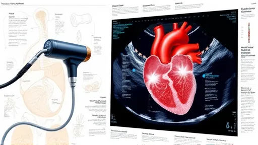
The study also raises a question that the authors do not fully address: what about the bubbles that are not visible at all? The TEE images show what the ultrasound can capture, but ultrasound has physical limits. Small bubbles, deeply located bubbles, bubbles moving at certain angles to the probe — these may simply not appear in the image, no matter how good the detection algorithm is. The model can only detect what the data contains. If the data is blind, the model is blind too, and no amount of architectural sophistication can change that.
This is the fundamental limitation that all AI systems share, and it is worth stating plainly: the algorithm is only as good as the data it sees, and the data is always incomplete. The model does not know what it is missing. It does not have a sense of its own blind spots. It produces output with the same confidence regardless of whether the input is perfect or degraded. A human expert, by contrast, knows when the image quality is poor, knows when the view is inadequate, knows when to say “I cannot tell.” The model has no such self-awareness, and the grace zone evaluation does not reward self-awareness. It rewards the confident production of masks, even when those masks are wrong.
The Cost of Convenience
There is a seductive logic to AI-assisted medicine that is hard to argue against. The model is fast, it is tireless, it does not get distracted, it does not have a bad day. All of this is true. But the same logic applies to a spellchecker, and we all know that a spellchecker cannot write a sentence. The tool is useful precisely because it handles the mechanical parts, freeing the human to focus on the parts that require judgment. The danger arises when the mechanical parts and the judgment parts become blurred, when the tool’s output is treated as if it were judgment itself.
In the cardiac operating room, the stakes are unusually high. The brain is perfused by the heart, and every bubble that crosses the arterial system is a potential insult to that brain. The study’s title — “Protect the Brain When Treating the Heart” — captures the clinical imperative perfectly. But the model does not protect the brain. It provides information that a human might use to protect the brain, and the quality of that information is variable in ways that are not always visible. The grace zone makes the information look better than it is. The strict metrics reveal the truth. The question is which version of the truth will be presented to the surgeon when this system moves from feasibility study to clinical deployment.
The Uncomfortable Question
The paper ends with the standard conclusion: the results support the technical feasibility of real-time GME segmentation. This is a modest claim, and it is probably true. The model can segment bubbles in real time, at least on the data it was trained on, at least under the evaluation conditions the researchers chose. But feasibility is not the same as readiness, and the gap between the two is where the real work remains to be done. The model needs to be tested on more patients, more machines, more conditions. It needs to be integrated into the clinical workflow in a way that supports rather than supplants human judgment. And it needs to be evaluated under strict metrics, not just the forgiving ones that make it look good.
The deeper question, the one that this study raises without answering, is whether we are willing to accept the kind of AI we are building. We are building systems that are fast, confident, and often imprecise. We are building systems that cannot explain themselves and cannot recognize their own limitations. We are building systems that perform well under conditions we choose and poorly under conditions we do not. And we are doing all of this because the alternative — doing the work ourselves, with our slow and fallible human attention — feels like too much to ask.
The Moment of Clarity
The surgeon in the operating room does not have the luxury of philosophical reflection. There is a patient on the table, a heart that needs fixing, a brain that needs protecting. The monitor shows the ultrasound feed, and somewhere in that feed there may be bubbles that the eye cannot catch. The algorithm is running, producing its masks, offering its confident approximations. The surgeon must decide what to do with that information, knowing that the model is not as precise as it appears, knowing that the grace zone was a choice, not a law of nature.
This is the moment where AI deception becomes concrete: not a lie, but a gap. The model claims to see, and it does see, but it sees in a way that is both more and less than human vision. It sees more because it processes every frame, every pixel, without fatigue. It sees less because it does not know what it is looking at, does not understand what a bubble means, does not carry the weight of a patient’s future in its calculations. The mask is empty or full, and the model does not care which. The surgeon cares, and that difference is the whole story.
The study is a small one, eight patients, a pilot, a feasibility check. But it is a window into a much larger pattern. We are building AI systems that look like they are solving problems while actually relocating them. The problem of detecting bubbles becomes the problem of trusting the detector. The problem of seeing becomes the problem of knowing what the seer is missing. And the grace zone, that small tolerance for error, becomes the space where we hide from the truth about our own creations. The model is not deceiving us. We are deceiving ourselves, and the algorithm is just the mirror we hold up to our own wishful thinking. The next time a surgeon glances at a mask and trusts it, the question is not whether the model is right — it is whether we have built a system that lets us avoid asking that question at all.