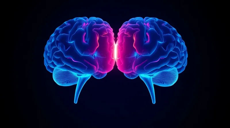
Two Ancient Brains Fused Into One
A Blueprint Written Twice, Not Once
That decision is not a single act of construction. It is two acts, running side by side, each with its own instruction set.
For as long as anatomists have drawn the nervous system, they have drawn it as one thing. A single sheet of tissue rolls into a tube. That tube swells at one end. The swellings become the forebrain, the midbrain, the hindbrain — one organ, three neighborhoods, one origin story.
A study led by developmental biologists at Stanford Medicine, published in Nature Neuroscience, now overturns that picture. What we call the human brain, the authors argue, is not one structure that diversified. It is two ancient nervous systems that evolution pushed together and wired into a single skull.
The distinction matters because it is not a matter of degree. It is a matter of kind. The front of the brain and the back of the brain do not share a common progenitor cell. They never did.
Kyle Loh, Ph.D., associate professor of developmental biology at Stanford Medicine and senior author of the study, put the finding in plain terms: the front of the brain arises from a totally different progenitor cell than the back of the brain
The adult brain still divides into three classical regions. The forebrain handles executive cognition, language, and abstract thought. The hindbrain — what most people call the brainstem — runs the machinery no one thinks about: rhythmic breathing, cardiac regulation, sleep-wake cycles, metabolic drive, and the motor control behind facial expression, speech, and swallowing. These two domains, it turns out, were never siblings. They were strangers assigned to the same address.
The Lock Inside the Earliest Cells
The obstacle that stalled brainstem research for decades was never a lack of effort. It was a lock. The Stanford team found that lock by looking at the earliest stage of mouse embryogenesis — gastrulation — when the embryo is still a disk of cells deciding what to become.
Co-first authors Rayyan Jokhai and Carolyn Dundes identified two progenitor populations that never overlap. One expresses a gene called Otx2. It is dedicated exclusively to the forebrain and midbrain. The other expresses Gbx2. It is committed from the outset to the hindbrain. These two cell populations do not cross lineages. They do not trade cells. They do not merge.
The reason they cannot merge is physical. Epigenomic evaluations revealed that the anterior and posterior neural ectoderm carry fundamentally different chromatin packaging architectures. Chromatin is the material that wraps DNA and decides which genes a cell is allowed to read. In these two cell types, that packaging is configured differently from the very start, and it locks each cell into its trajectory permanently.
This explains a long history of failure. Previous attempts to make hindbrain neurons likely tried to coax forebrain and midbrain progenitors into hindbrain cells, which the work shows is not possible. The cells were not stubborn. They were already committed to a different fate before anyone tried to redirect them.
Jokhai drew a larger lesson from this. In stem cell biology, he noted, researchers are always fixated on creating the end cell type — the neuron itself. But it is important to begin at the earliest stages of embryonic development. Attention to that early moment is what exposed the split. Skip it, and a foundation is built that cannot bear the load.
The practical consequence is concrete. By respecting the early split, the Stanford investigators guided human pluripotent stem cells into fully functional hindbrain motor neurons. The lab-grown cells displayed normal action potentials — the electrical spikes neurons use to signal — and expressed markers characteristic of the hindbrain segments that control facial musculature and swallowing. These are the first authentic human hindbrain neurons grown in a dish.
Two Nervous Systems, Pushed Together
To trace where this division came from, the researchers looked far beyond mammals. They found the same dual-origin mechanism in other vertebrate species. More primitive organisms carry two separate nervous systems positioned at opposite ends of their bodies.
That pattern suggests a sequence. Two nervous systems existed first, apart. Then evolution brought them together. Loh described the logic: evolution took two existing neural systems and pushed them together spatially. He added a note that cuts against intuition — having the brain as one organ would probably be more efficient, yet we rely on this primordial way of making the brain as two separate pieces.
The clinical reach of this work is direct. In amyotrophic lateral sclerosis and spinal muscular atrophy, degeneration of hindbrain motor neurons takes away a patient’s ability to swallow and breathe. Research has been stalled because living brainstem tissue cannot be taken from patients. There has been no accessible human cellular model — until now.
Jokhai framed what this opens: there is now a model to better understand these devastating diseases and to work toward regenerative therapies for them. He called it a new frontier in brain research. The frontier is not a metaphor. It is a dish of living human neurons that, for the first time, come from the hindbrain half of the brain’s ancient, two-part inheritance.
Sources
1. Nature Neuroscience (2026-09-18) — DOI
2. Medicalxpress — Quote source (original article)