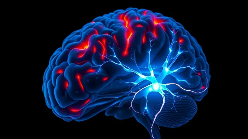
Stroke triggers flawed growth of new brain neurons
A Systematic Look Reveals a Consistent, Flawed Response
For years, scientists have known that a stroke triggers the birth of new neurons in the adult hippocampus, a brain region critical for memory and learning. Earlier studies, however, often relied on a single laboratory model of stroke. That approach left an open question: Is the observed response a general biological rule, or just an artifact of one particular experimental setup? A new multicenter study, conducted within the Stroke-IMPaCT consortium across six different sites, set out to answer that question by applying a systematic, multimodel analysis for the first time.
The consortium used two main types of stroke models in adult C57BL/6J mice: permanent and transient middle cerebral artery occlusion. Within those categories, they employed a distal middle cerebral artery occlusion under normal oxygen conditions, the same occlusion under low oxygen (hypoxia), and a filament-based transient occlusion. By comparing results across these very different methods of inducing ischemia, the researchers could isolate which cellular changes are truly conserved and which are specific to a given injury type.
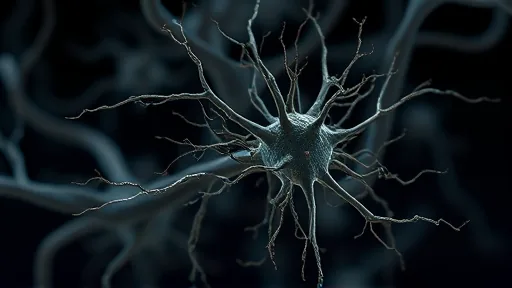
The findings were stark and uniform. Across every model and every site, the mouse hippocampus responded to stroke with a sharp, bilateral increase in cell proliferation — measured by the marker Ki67. This burst was strongest at three days after the stroke and remained elevated at seven days. By two months, the proliferation rate had returned to baseline. The number of neuroblasts, young neurons marked by doublecortin (DCX), also increased, particularly on the side of the brain directly affected by the stroke, but this, too, normalized over time.
The Quality of New Neurons, Not Just Their Quantity, Is the Problem
While the initial increase in cell numbers might seem like a positive, regenerative response, the long-term analysis told a different story. The researchers performed high-resolution examinations of the newborn neurons’ structure, focusing on their dendritic architecture and somatodendritic polarity — essentially, the shape and orientation of the cell’s receiving branches. What they found was consistent across all models: the new neurons were structurally abnormal.
Specifically, the apical dendrites — the main branches that receive input from other neurons — were significantly shorter than normal. More troubling, a large proportion of these newborn cells displayed what the study calls “aberrant features.” These include ectopic positioning (the neuron is in the wrong place), polarity defects (the cell’s internal organization is scrambled), and abnormal lateral growth (branches grow sideways instead of upward). These are not subtle variations; they are fundamental architectural errors that prevent the neuron from connecting properly into the existing brain circuit.
This finding reframes the entire understanding of post-stroke neurogenesis. The brain does produce new cells, but the cells it produces are structurally compromised. The quality of newborn neurons, not just their quantity, is the critical factor A brain full of misplaced, malformed neurons cannot compensate for lost function. This structural deficit may be a direct contributor to the long-term cognitive problems — including dementia and cognitive dysfunction — that many stroke survivors face.
A Conserved Hallmark That Points Toward Future Therapy
The final and most anchoring data point from the study is the conclusion that this aberrant hippocampal neurogenesis is a “robust hallmark of poststroke pathology in mice.” The word “robust” is chosen deliberately. It means the finding is not fragile or dependent on a specific lab technique. It holds true whether the stroke is permanent or temporary, whether it involves a large filament or a small distal occlusion, and whether the mouse breathes normal air or low oxygen. The response is conserved.
This conservation has a profound implication for future research and therapy. If the malformed neuron response is a universal consequence of stroke, then any treatment designed to enhance brain repair must address this structural quality issue. Simply boosting the number of newborn cells is unlikely to help; in fact, it might worsen the problem by filling the hippocampus with more dysfunctional units. The study explicitly underscores the need to evaluate “structural quality” when developing therapeutic strategies.
The mechanism driving this flawed development remains unknown, as the study is observational, not interventional. But by establishing that the phenomenon is a conserved, model-independent response, the Stroke-IMPaCT consortium has provided a clear and stable target. The next step is to investigate the molecular signals that guide a newborn neuron’s migration and dendrite growth in the post-stroke environment Only by understanding what goes wrong can scientists begin to design ways to correct it.