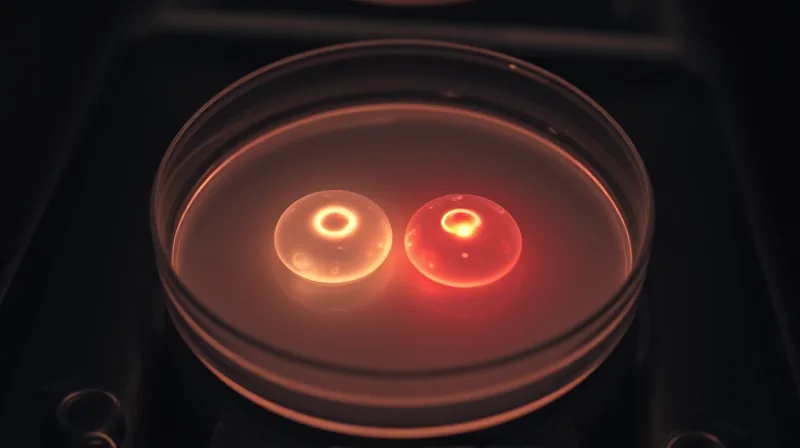
Lab oxygen mistake reveals IVF embryos need 2 percent oxygen
The Mistake That Rewrote the Playbook
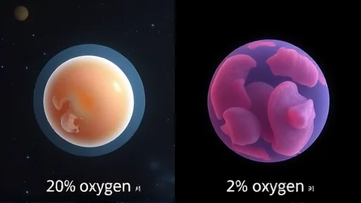
For decades, embryologists in IVF clinics grew embryos in an environment that was, in hindsight, far too rich in oxygen. The standard practice, born from the convenience of atmospheric air, exposed developing embryos to roughly 20% oxygen. This was not a deliberate choice based on biology. It was a default setting. Lab incubators were simply set to match the air we breathe. The mistake was hiding in plain sight: the inside of a human body is not an open field. It is a low-oxygen world.
The reproductive tract of a mammal is a journey of falling oxygen. In the fallopian tubes, where fertilization occurs, oxygen levels hover around 7%. By the time an embryo reaches the uterus, that number drops to just 2%. The developing cells are designed for this gradient. They are adapted to scarcity. Marisa Bartolomei at the University of Pennsylvania explains that “historically, people would do IVF at atmospheric oxygen levels — about 20 per cent.” [2] This mismatch was not a minor detail. It may have been a fundamental flaw in how embryos were grown outside the body.
The consequences of this oversight are now becoming clear. Current guidelines for IVF clinics recommend culturing embryos at 5% oxygen. This lower level helps limit damage from oxidative stress — an imbalance of free radicals and antioxidants that can harm cells. But even 5% is still more than double the oxygen concentration an embryo would encounter inside the uterus. Some researchers suspected that dropping the level even further, to 2%, might be better. The question was whether this adjustment would make a measurable difference in the health of the resulting offspring.
Bartolomei and her team decided to test this hypothesis directly. They cultured mouse embryos at two different oxygen concentrations: one group at 2% oxygen, the other at the current standard of 5% oxygen. After this lab phase, they transferred the embryos into the uteruses of female mice, exactly as is done in routine IVF. This was not a short-term experiment. The researchers followed the offspring for 12 weeks after birth. Amy Sparks at the University of Iowa, who was not involved in the research, calls this a “landmark paper that we will cite for decades to come.” [3] She notes that most studies on oxygen concentration “have stopped at the termination of embryo culture. This goes so far beyond.”
The results were striking. The embryos cultured at 2% oxygen more closely resembled naturally conceived embryos than those grown at 5% oxygen. The similarities were not just superficial. They extended to the level of epigenetic reprogramming — the process by which chemical tags on DNA switch genes on or off. This reprogramming is essential for an embryo to develop properly before it implants in the uterus. Many of the enzymes that perform this delicate work are sensitive to oxygen. When oxygen is too high, these enzymes may become overactive. “Our idea is that they overshoot the mark,” says Bartolomei. “They become too active.”
The Timeline From Mouse to Human Clinic
The path from this discovery to a change in human IVF practice is not short. The first step, already taken, is the mouse study itself. The next step, according to researchers, must involve larger animals. Nina Boskovic at the Karolinska Institute in Stockholm, Sweden, specifically recommends testing in cows and pigs. [4] Their reproductive biology is closer to that of humans. If the results hold in these animals, then and only then can human trials begin.
The safety testing is non-negotiable. The mice in the 2% oxygen group showed no known side effects from the intervention. But what works in a mouse does not always work in a person. The human trials must test both safety and efficacy. Embryos are delicate. Changing the oxygen level changes the entire chemical environment in which they develop. Before any clinic adopts a new protocol, the evidence must be solid.
If the human trials are successful, the technical adjustment is straightforward. Bartolomei estimates that clinics would need to change their incubators and their gas supplies. This is not a trivial expense, but it is small compared to the overall cost of IVF. “It’ll be costly in the sense that they have to change their incubators and gases,” she says. “But compared to how much IVF costs, it really isn’t very costly.” The timeline for this transition depends entirely on the speed of the research pipeline.
This is not the only avenue of research aimed at improving IVF outcomes. A separate line of work, published on arXiv, focuses on using artificial intelligence to predict which embryo is most likely to implant successfully. One study from June 2020, titled “Data-Driven Prediction of Embryo Implantation Probability Using IVF Time-lapse Imaging,” trained an algorithm on time-lapse videos of 272 embryos. The algorithm outperformed a panel of human embryologists. It achieved a 12% increase in positive predictive value and a 29% increase in negative predictive value. This work addresses a different problem — selection — but it shares the same ultimate goal: making IVF more successful.
Another paper from October 2024, “Multimodal Learning for Embryo Viability Prediction in Clinical IVF,” takes this approach further. It combines time-lapse video data with electronic health records to predict embryo viability. The challenge here is fusing two very different types of data: moving images of cells dividing and static medical histories. The researchers are developing a model that can handle both. This automated system could replace the subjective, manual evaluation that embryologists currently perform. It is a parallel effort to reduce the guesswork in IVF.
The Final Data Point That Anchors the Finding
The study from Bartolomei’s team provides a specific, measurable outcome. Embryos cultured at 5% oxygen had more trophectoderm cells at the blastocyst stage. These cells are important — they help form the placenta. But too many of them is a problem. An excess of trophectoderm cells may explain why the placenta can be thicker in IVF pregnancies. And a thicker placenta has been linked to a lower birth weight.
The data from the mice confirmed this link. The offspring from the 5% oxygen group had a lower birth weight than those from the 2% oxygen group. This is not a subtle difference. It is a direct, observable consequence of the oxygen level during the earliest days of development. The 2% group produced offspring that were closer in weight to naturally conceived mice.
The number of babies born via IVF in the UK has risen sharply. In the year 2000, it was about 8,700. By 2023, that number had grown to 20,700. IVF is a safe and well-established procedure. But it carries a higher risk of low birth weight. This study offers a potential explanation for that risk. It also offers a path toward reducing it. .
The mechanism is not yet fully understood. Bartolomei proposes two possibilities. Lower oxygen levels might allow embryos to use energy in a way that more closely resembles development inside the uterus. Alternatively, the effect might be mediated through epigenetic reprogramming. The enzymes involved in resetting gene activity are sensitive to oxygen. At higher levels, they may become too active and overshoot their target. The result is a cascade of changes that affect the placenta and, ultimately, the baby’s birth weight.
The study is published on bioRxiv with the DOI 10.64898/2026.07.10.737757. It is a preprint, meaning it has not yet undergone peer review. But the data is already prompting discussion. Sparks hopes the study ‘will encourage others to do quite a bit of research in this area.’ The final data point is clear: a 2% oxygen environment produces healthier mouse embryos. The question now is whether the same holds true for humans.
Sources
1. DOI: 10.64898/2026.07.10.737757