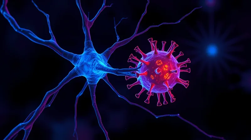
Epstein-Barr Virus Triggers Multiple Sclerosis Immune Attack
The Old Model Could Not Explain the Trigger
For years, the story of multiple sclerosis had a gap at its center. Doctors knew that the immune system attacked the protective myelin sheath around nerves in the brain and spinal cord. They could see the damage on scans. They could measure the worsening symptoms: vision loss, difficulty walking, numbness. But they could not answer the simplest question: why did the immune system turn against the body in the first place?
A common virus, Epstein-Barr virus, had been a suspect for decades. The virus infects about 90 percent of the global population. For most people, it causes nothing more than a brief illness — sometimes not even that. Yet virtually everyone with multiple sclerosis carries the virus. In 2022, a study of more than 10 million people found that the risk of MS is 32 times higher after an Epstein-Barr virus infection. [1] No other viral connection was found. The correlation was overwhelming. But correlation is not mechanism. The old model could show that the virus was present. It could not show how the virus made the immune system attack the brain.

The Mechanism That Connects Virus to Immune Attack
The new research, published in Science Translational Medicine, began with a simple measurement. Researchers led by Kjetil Bjornevik at the Harvard T.H. Chan School of Public Health and Natalia Drosu at Massachusetts General Hospital compared immune T-cell activity in people with MS and in healthy control individuals. [1] They found that T-cell activity was twice as high in the MS group. That was a clear difference. But it was also a crude measurement. The immune system contains many types of T cells. Which ones were driving the response?
The researchers then selectively depleted various kinds of T cells from the participants’ blood samples. When they removed a type called CD4+ T cells, the immune response to Epstein-Barr virus dropped sharply. The CD4+ T cells were the drivers. That was a specific finding. But it raised a new question: how were the CD4+ T cells being activated? The researchers knew that another type of immune cell, called B cells, can harbor the Epstein-Barr virus. They also knew that a class of drugs called anti-CD20 therapies works by destroying B cells. But the mechanism connecting B cell destruction to reduced T-cell activity was unclear.
To find out, the researchers measured CD4+ T-cell levels in 60 people with MS before and six months after starting an anti-CD20 treatment. The levels had decreased by about 2.5 times. The team validated the results in a second group and found that the reduction persisted for up to a year. Another group of people who received anti-CD20 treatment had lower levels of Epstein-Barr virus in their saliva than healthy people or people with untreated MS. That suggested that the drug reduced viral activity. But the researchers could not directly observe the moment when a B cell released a viral particle that activated a CD4+ T cell. That step remained a black box. The technical limits of current imaging and sampling methods meant that the precise molecular handshake between the infected B cell and the T cell could not yet be filmed or measured in real time. The researchers could see the before and after. They could not see the middle.
The Parallel Finding That Confirms This Line of Research
While the Harvard and Massachusetts General team worked on CD4+ T cells, a separate group of researchers at the University of Texas Health Science Center pursued a related question. Assaf Gottlieb and colleagues wanted to know which T cells were present in the cerebrospinal fluid of people with early symptoms of MS — before they had received a formal diagnosis. They sequenced the receptors on the outside of T cells to see what the cells recognized.
The results were striking. In the patients’ blood samples, 13 percent of T cells had receptors recognizing Epstein-Barr virus-infected cells. Only 4 percent recognized antigens for the flu. In the cerebrospinal fluid, the proportion of T cells recognizing Epstein-Barr virus-infected cells jumped to 47 percent. These were not just any T cells. They were clones — copies of the same T cell that had multiplied in response to the virus. The finding came from a small study of only eight patients. But it complemented the larger study in a crucial way. The cerebrospinal fluid bathes the brain and spinal cord. Finding virus-specific T cells there, at the earliest stage of the disease, strongly suggested that these cells were not bystanders. They were either causing the damage or contributing to it.